

“Ovary Preserving Hysterectomy” Illustration by Abigail Rumpp
Ovary Preserving Hysterectomy
Year
2025
Content Expert
Rebecca Stone, MS, MD
Jeni Fairman, MA, MPS, CMI, FAMI
Medium
Photoshop, graphite
About
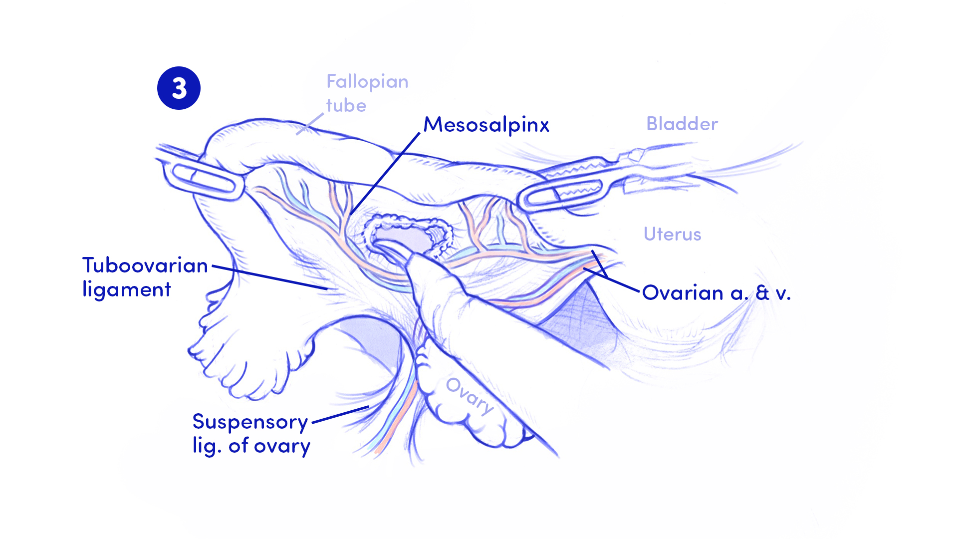
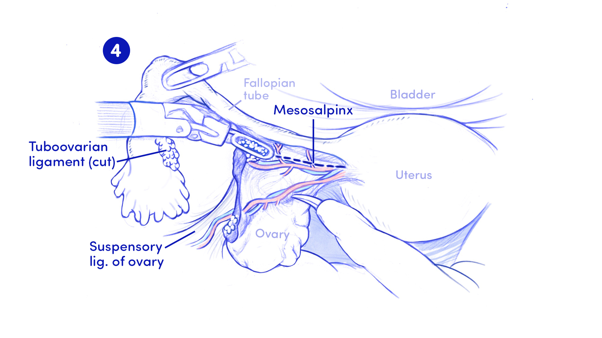
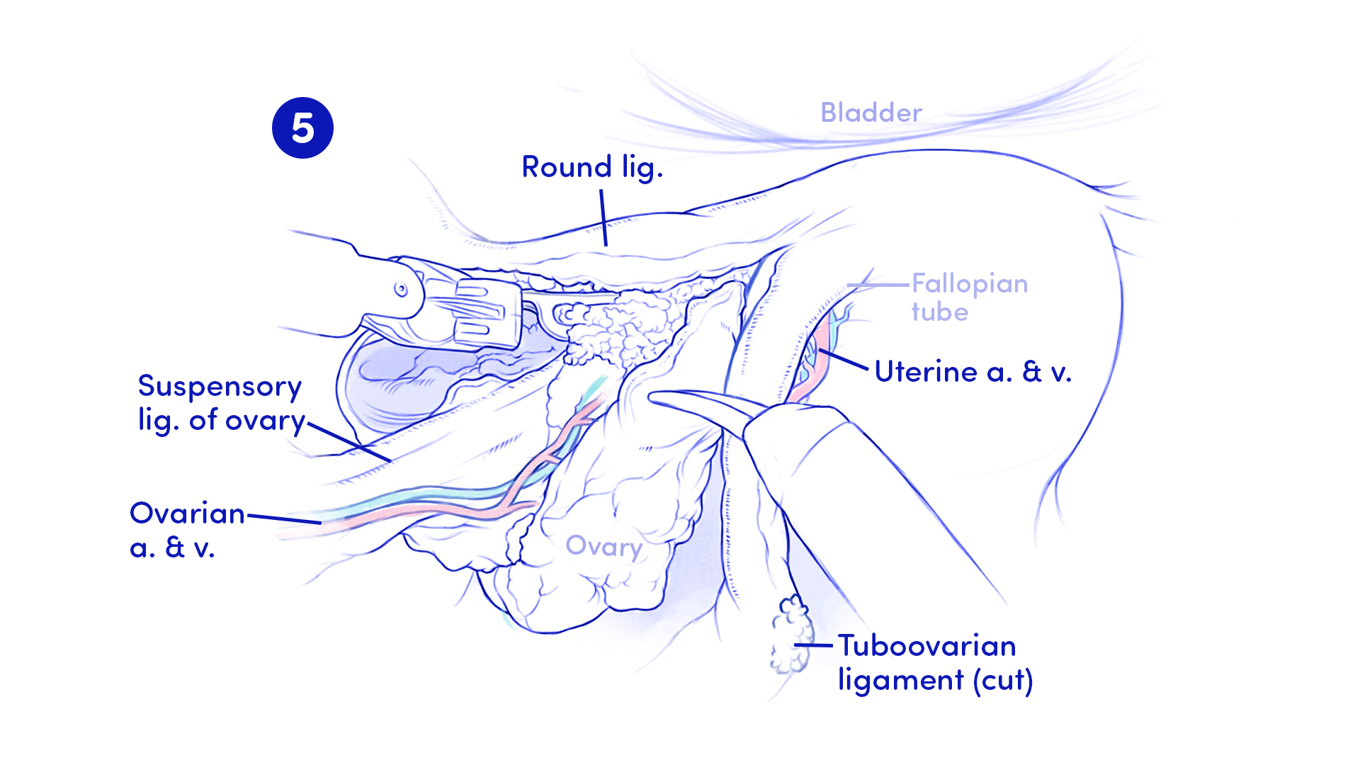
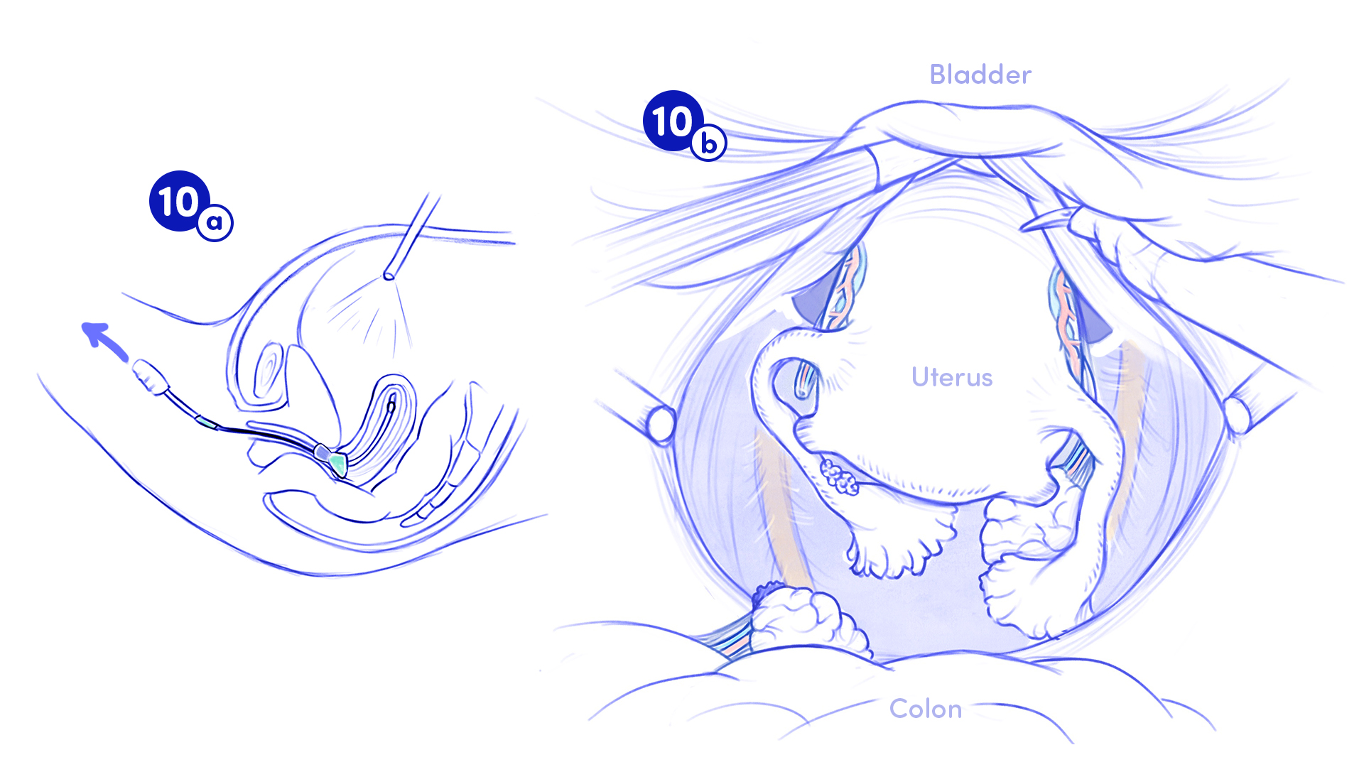
This illustration was created following the observation of a laparoscopic ovary-preserving hysterectomy performed by Dr. Rebecca Stone at The Johns Hopkins Hospital. It depicts a critical step in the procedure: the division of the mesosalpinx using bipolar forceps, with careful dissection along the ovarian artery and vein to preserve the ovary’s blood supply. This technique is essential to ensure the ovary remains functional and perfused postoperatively.
The illustration was created for an audience of OB-GYN surgeons and residents.

The surgeon follows the ovarian artery and vein as the mesosalpinx is divided in order to keep the ovary perfused.
Background
A hysterectomy is a surgical procedure in which the uterus is removed. In a total hysterectomy, both the uterus and cervix are removed. Depending on the patient’s needs or preferences, other structures, such as the fallopian tubes and ovaries, may also be removed.
In the surgery I observed, the patient elected to preserve their left ovary. Retaining even one ovary can help delay the onset of menopause, as it continues to produce the hormones estrogen and progesterone. This is often preferred for younger patients or those with health conditions that could be worsened by the sudden loss of these hormones.
Illustrated Surgical Steps
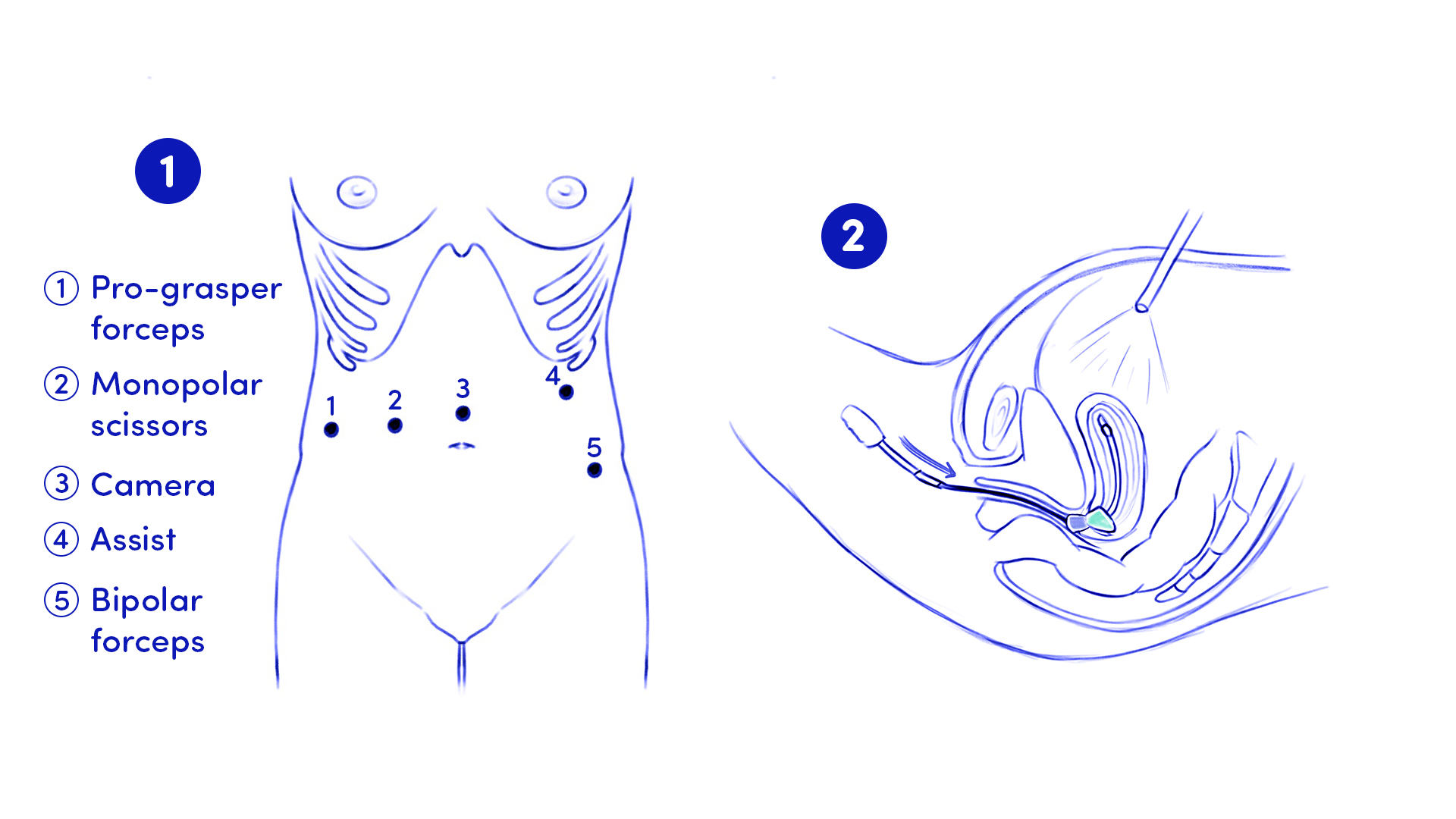
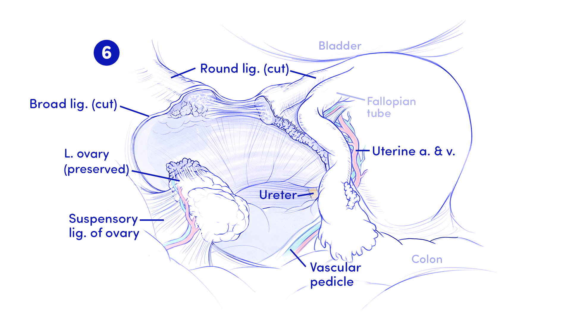
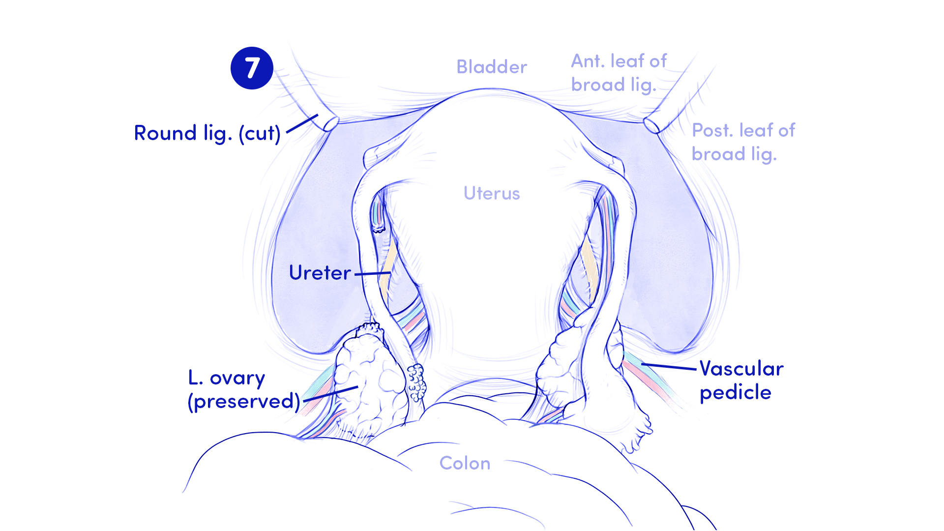
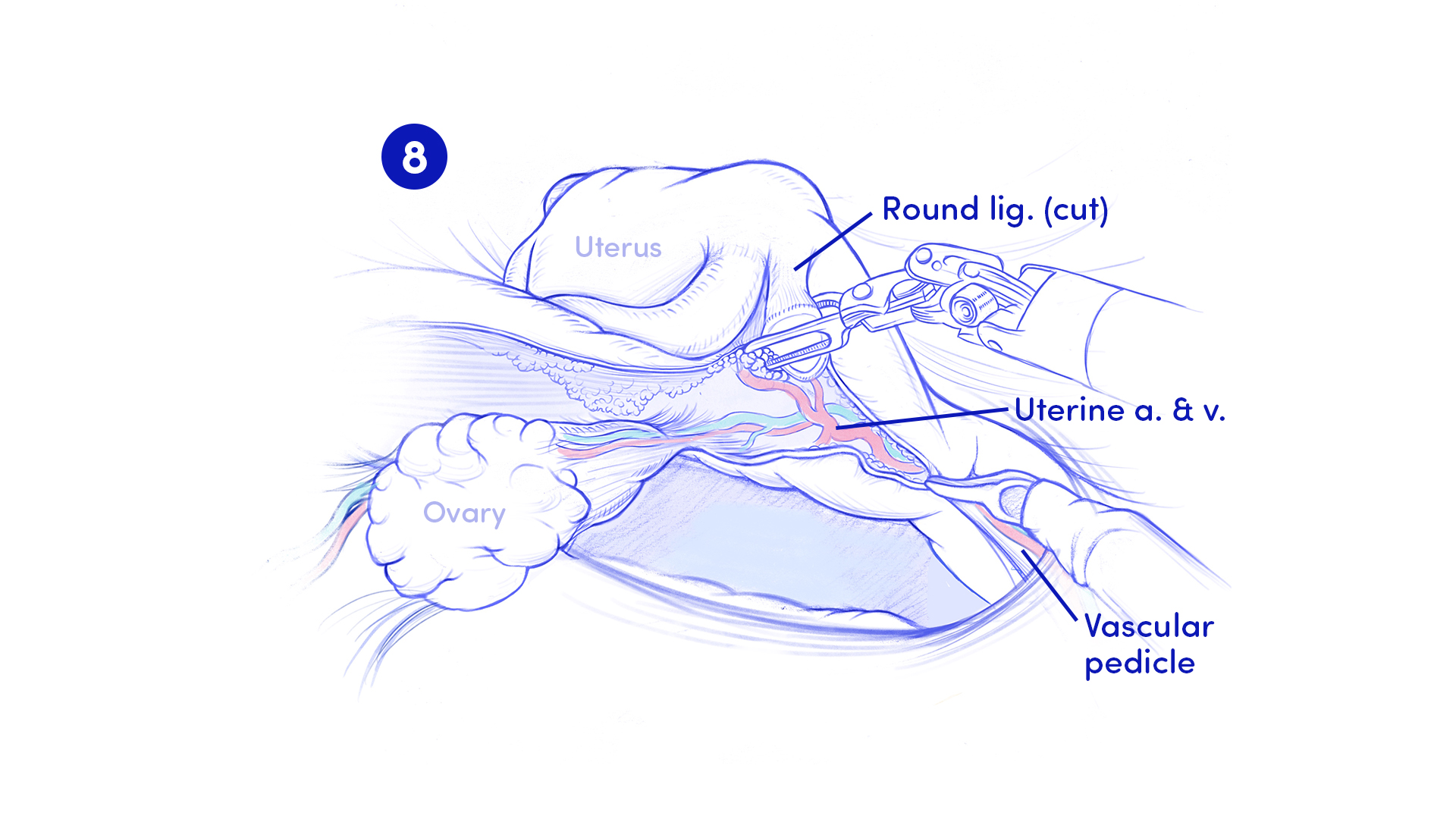
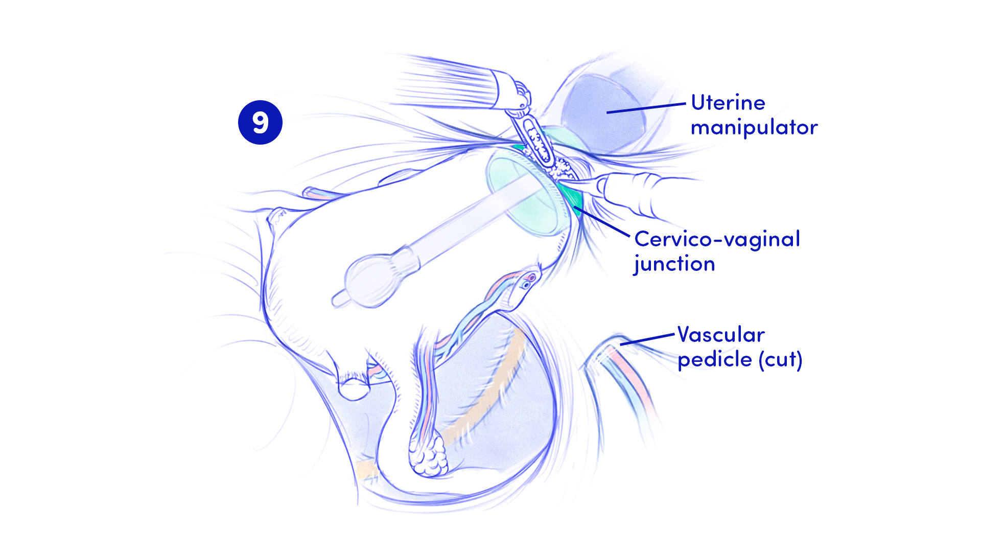
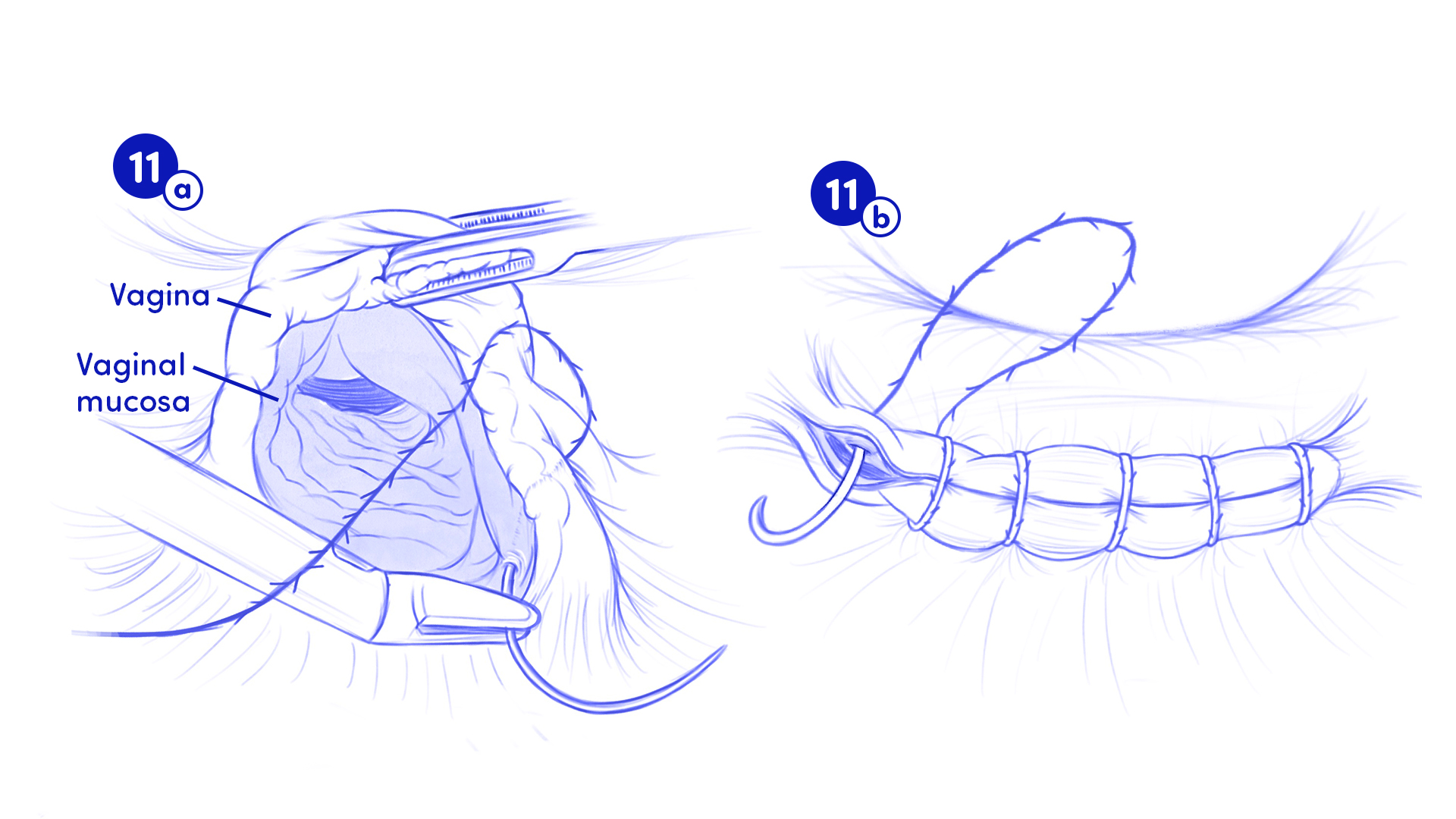
OR sketches of the surgical steps observed during the procedure.